Nervous-system questions punish school biology harder than almost anything else on the USABO. Your syllabus taught you that a wave travels down an axon; the exam asks which ion moved, in which direction, driven by what, and what happens if you block one gate. This is the quantitative core, and the four calculations that turn it from memory into method.
Where these questions live, and why they feel disproportionately hard
Animal anatomy and physiology is the largest content band on the exam, and our own reading of the material puts it at roughly 25%. Nervous-system material sits inside that band. CEE publishes the content outline, so confirm the current version on cee.org before you plan around any particular sub-topic. If you want the format context first, our overview of the USABO covers the structure this article assumes: 50 questions in 50 minutes, no penalty for a wrong answer, roughly sixty seconds a question.
The difficulty is not that neurophysiology is intrinsically harder than immunology or plant transport. It is that the gap between your syllabus and the exam is unusually wide here, in a specific way. AP, IB and A-Level all teach the action potential as a narrative — depolarisation, repolarisation, refractory period, done. The olympiad treats the same material as electrochemistry: concentrations, equilibrium potentials, driving forces, conductances and time constants. A narrative cannot answer a question that gives you two ion concentrations and asks which way current flows.
The good news is that this band is unusually learnable. Three ideas — selective permeability, two gates on one channel, and driving force — generate the answers to most of what gets asked. Learn those properly and the rest is arithmetic.
The resting potential is a permeability argument, not a pump argument
Ask a school-taught student why the inside of a neuron is negative and you will usually hear “the sodium-potassium pump.” That is the most common half-right answer on this topic, and the exam knows it.
The pump (three Na+ out, two K+ in, per ATP) builds and maintains the gradients. Its direct electrogenic contribution to the voltage is small. The voltage itself comes from selective permeability: at rest the membrane is far more permeable to K+ than to Na+, so the membrane potential sits close to the potassium equilibrium potential, pulled a little positive by a small persistent Na+ leak.
The equilibrium potential for any ion comes from the Nernst equation. At body temperature, using base-10 logarithms, a workable form is:
Eion ≈ (61.5 / z) × log10 ( [ion]outside / [ion]inside ) mV
where z is the ionic charge. The constant is temperature-dependent (you will see roughly 58 to 62 quoted depending on the temperature assumed), so read what the question gives you rather than reciting a number.
| Ion | Typical outside | Typical inside | z | Eion | What this means at rest (Vm ≈ −70 mV) |
|---|---|---|---|---|---|
| K+ | 5 mM | 140 mM | +1 | about −89 mV | Vm sits near here. Small outward driving force, so K+ trickles out |
| Na+ | 145 mM | 15 mM | +1 | about +61 mV | Huge inward driving force, but permeability is low — so almost nothing happens until channels open |
| Cl− | 110 mM | 10 mM | −1 | about −64 mV | Close to rest, so opening Cl− channels often clamps rather than hyperpolarises |
| Ca2+ | 2 mM | ~100 nM | +2 | about +132 mV | The steepest gradient in the cell — which is why Ca2+ works so well as a signal |
One asymmetry in that table answers a whole family of exam questions. Raise external K+ and the resting potential moves substantially (because Vm tracks EK, and EK is sensitive to the outside concentration when it is small). Change external Na+ and the resting potential barely moves, but the peak of the action potential collapses (because the peak tracks ENa). If a stem manipulates an external concentration, that single contrast usually tells you which part of the trace should change.
The action potential, and the two gates that explain everything
Almost every action-potential question reduces to one structural fact: the voltage-gated sodium channel has two gates, not one. An activation gate that opens fast on depolarisation, and an inactivation gate that closes more slowly and cannot reset until the membrane repolarises.
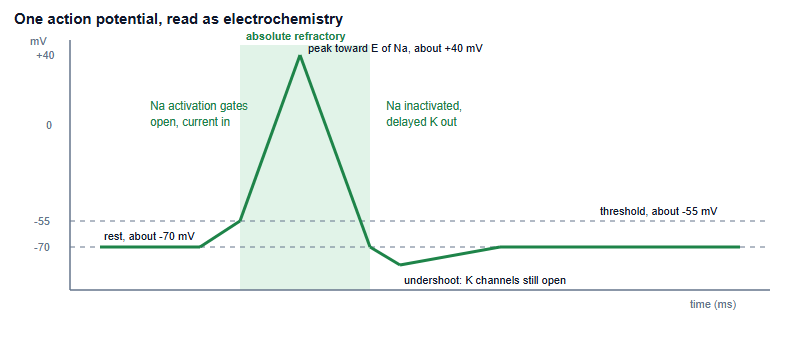
That one structural fact generates five separate exam answers:
- Threshold exists because Na+ entry is self-reinforcing: entry depolarises, depolarisation opens more channels. Below threshold, K+ leak wins; above it, the loop runs away.
- All-or-none follows — once the loop runs, amplitude is set by the ion gradients, not by how hard you poked it. Stimulus intensity is therefore coded by firing frequency and by how many fibres are recruited, never by spike height. That is a standard distractor.
- The absolute refractory period is inactivation gates being shut. No stimulus of any size works, because the channels are physically unavailable.
- One-way propagation follows from the same thing: the membrane behind the spike is inactivated, so current spreading backwards cannot re-fire it.
- Maximum firing rate is set by the refractory period. If the absolute refractory period is about 2 ms, the ceiling is about 1000 / 2 = 500 impulses per second. Exams ask exactly this.
Add one more idea and you can explain the shape rather than recite it: driving force = Vm − Eion. At rest, the driving force on Na+ is enormous (−70 minus +61, roughly −131 mV of inward push) but permeability is tiny, so little flows. At the peak, the Na+ driving force has nearly vanished while the K+ driving force is at its largest — which is precisely why repolarisation is so fast. Current is always conductance multiplied by driving force, and questions that look impossible usually just want you to notice which of those two terms changed.
Conduction speed: two solutions to one problem
Between spikes, signal spreads passively, and it decays as it goes. How far it gets before decaying is set by the length constant, which improves when membrane resistance rises or internal resistance falls. Evolution has found two independent routes to a faster axon:
- Wider axons lower internal resistance, because a fatter cable carries current more easily. This is the invertebrate solution — the squid giant axon, the preparation the classic electrophysiology was done on.
- Myelin raises membrane resistance and lowers membrane capacitance, so current spreads further and charges the membrane faster, reaching the next node of Ranvier sooner. Conduction jumps node to node.
The comparative point is worth having ready: myelin buys speed far more cheaply in space and metabolic cost than diameter does, which is why a thin myelinated vertebrate axon can outrun a much wider unmyelinated one. And the clinical corollary is the exam-friendly half: strip the myelin and conduction slows or fails, because current leaks away before it reaches the next node.
The timing calculation. Latency questions are worth practising because they look like biology and behave like arithmetic. A signal covering 1 m at 50 m/s takes 20 ms. If the measured reflex latency is 24 ms, then about 4 ms is unaccounted for. Each chemical synapse costs roughly 0.5 to 1 ms of delay, so that gap implies several synapses — a polysynaptic pathway, not a monosynaptic one. Reasoning from an unexplained delay to a circuit is a classic olympiad move.
Synapses: the three questions every synapse item is really asking
Synaptic questions look varied and are not. Strip the scenario away and almost all of them ask the same three things in sequence.
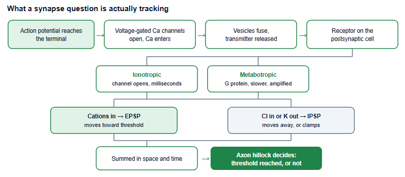
The single most useful correction to school biology here: excitation and inhibition are properties of the receptor, not of the transmitter. Acetylcholine excites skeletal muscle through a nicotinic receptor that lets cations in; the same acetylcholine slows the heart through a muscarinic receptor that opens K+ channels and hyperpolarises. Any question phrased as “is transmitter X excitatory?” is testing whether you know the question is malformed.
Summation is the other reliably examined idea. A single EPSP is usually far too small to reach threshold, so inputs must add — temporally (the same terminal firing repeatedly before the previous potential decays) or spatially (many terminals firing at once). The addition is judged at the axon hillock, where voltage-gated Na+ channels are densest. Inhibition placed close to the hillock can also work by shunting: opening Cl− channels near rest may barely change the voltage while still bleeding away incoming current.
Finally, learn the interference table. Exams probe mechanism by breaking one step and asking what survives — this is the highest-yield half-page in the whole topic.
| Agent | Target | What still works | What fails |
|---|---|---|---|
| Tetrodotoxin | Voltage-gated Na+ channels | Resting potential is roughly normal | No action potential can be generated or propagated |
| Tetraethylammonium | Voltage-gated K+ channels | Upstroke is normal | Repolarisation is slowed, so the spike is prolonged |
| Curare | Nicotinic acetylcholine receptor | Nerve conducts normally; transmitter is released | Muscle does not respond — the block is postsynaptic |
| Botulinum toxin | Vesicle fusion machinery | Nerve conducts; receptors are intact | No transmitter is released — the block is presynaptic |
| Anticholinesterase | Acetylcholinesterase | Release and binding both occur | Transmitter is not cleared, so the signal persists |
| Ouabain | Na+/K+ ATPase | Initial spikes still fire | Gradients run down over time, so firing eventually fails |
Notice the pattern the table is really teaching: curare and botulinum toxin both produce a limp muscle, but for opposite reasons — one postsynaptic, one presynaptic. A well-written stem gives you exactly one observation that separates them, such as whether transmitter is detectable in the cleft.
Practising it, and the counterintuitive results worth knowing cold
Four habits convert this material into marks. Redraw the action potential from memory once a week with both axes labelled and every phase annotated with the ion and its driving force. Do the arithmetic until it is automatic: one Nernst calculation, one driving-force sign, one refractory-period ceiling, one latency estimate. Convert every mechanism you meet into a perturbation question — if I block this, what changes and what does not — because that is the form the exam uses. And then test all of it under time, using the past-paper pack rather than untimed textbook questions, since sixty seconds is part of the difficulty.
Three results in sensory neurobiology are worth knowing precisely, because they are counterintuitive and therefore heavily used as distractors:
- Light hyperpolarises vertebrate photoreceptors. Light activates rhodopsin, which activates transducin, which activates a phosphodiesterase that destroys cGMP; cyclic-nucleotide-gated channels then close, cation entry stops, and the cell hyperpolarises and releases less glutamate. The stimulus reduces the signal.
- Receptor potentials are graded; action potentials are not. Intensity is encoded as frequency downstream, which is why a stronger stimulus produces more spikes rather than bigger ones.
- Adaptation is a feature, not a fault. Phasic receptors report change and fall silent under a constant stimulus; tonic receptors keep reporting. Which one a question describes usually determines the answer.
If you are working out when in your school year to attack this band, remember that the exam calendar differs by region — check the China-region pathway page and cee.org before you plan the sequence. Neurophysiology rewards a long, early run at it rather than a fortnight of panic, because the arithmetic only becomes fast with repetition.
Is neurobiology a separate section of the USABO?
No. Nervous-system material sits inside animal anatomy and physiology; check the current content outline published on cee.org.
How much maths does this topic need?
Logarithms for a Nernst calculation and simple division for timing questions. Confirm any calculator policy on cee.org.
What is the single highest-yield idea here?
The two gates of the voltage-gated sodium channel. They explain threshold, refractory periods and one-way propagation at once.
Which textbook chapters cover this properly?
The nervous-system and sensory chapters of a general biology text, then a neuroscience text for channel-level and synaptic detail.
This is an independent guide operated by Hanlin Education for China-based international-school students. We are not affiliated with, endorsed by, or sponsored by the Center for Excellence in Education (CEE). Exam format, content weightings, eligibility and registration are set by the organisers — please confirm current details on cee.org. Ion concentrations and membrane values quoted above are typical textbook figures used for worked examples, not exam data, and always follow the values a question supplies. Statements about which ideas are most heavily examined, and about how questions are typically framed, are our coaches' editorial judgement, not official guidance. If you spot an error, tell us and we will correct it within 7 working days.