Circulation and gas-exchange questions on the USABO are rarely about naming heart chambers. They sit inside animal anatomy and physiology, the largest content area in our own reading of the syllabus, and they ask you to read an oxygen curve, predict what a pressure change does, or explain why a fish gill extracts oxygen so well. Four ideas carry almost all of them: partial-pressure gradients, the haemoglobin curve and its shifts, pressure and flow, and countercurrent exchange.
Where these questions sit, and what they are really asking
In the content weighting this guide works from, animal anatomy and physiology is the largest area at 25%, and circulation and gas exchange sit inside it; confirm the current content outline on cee.org. Our overview of the USABO covers the format assumed here: 50 questions in 50 minutes, with no penalty for a wrong answer.
At about a minute per question, these items come in three shapes. Curve items give you a dissociation curve or a pressure trace to read. Perturbation items change one variable, such as more carbon dioxide, a leaking valve or thinner air, and ask what follows. Comparative items describe an unfamiliar animal and ask which design suits its habitat. School courses teach labels — artery and vein, systole and diastole — while these items test the causes underneath them. In our coaching, the commonest wrong turn is answering with a label when the stem is really asking about a gradient.
Start with gradients: partial pressure, and why water is harder to breathe than air
Gases diffuse down partial-pressure gradients. Partial pressure is a gas’s fraction of the mixture multiplied by the total pressure, so dry sea-level air, about 21% oxygen at 760 mmHg, has an oxygen partial pressure (PO2) of roughly 160 mmHg. In the alveoli it is closer to 100 mmHg, and blood returning from resting tissues arrives at about 40 mmHg. Treat these as typical textbook values for sanity checks, and always work with the numbers a question gives.
Diffusion follows Fick’s law: the rate rises with surface area and with the partial-pressure difference, and falls as the barrier thickens. Emphysema shrinks the area, fibrosis thickens the barrier, and altitude shrinks the difference. High-altitude air is still about 21% oxygen, but total pressure falls, to roughly half its sea-level value near 5,500 metres, so an option claiming a lower oxygen percentage is a distractor. Carbon dioxide crosses readily despite a small gradient, roughly 46 against 40 mmHg, because it is about twenty times more soluble than oxygen.
Water is a harder medium. Even fully aerated, it holds roughly thirty times less oxygen per litre than air, oxygen diffuses thousands of times more slowly through it, and moving it across a surface is costly because it is far denser and more viscous. That is why gills depend on one-way flow and countercurrent exchange. It is also why mammals adjust breathing mainly in response to carbon dioxide and pH, whereas many water-breathers respond mainly to falling oxygen.
The oxygen curve: read the shape first, then the shifts
Haemoglobin (hemoglobin in American texts) has four oxygen-binding sites that cooperate: once one site binds, the next binds more readily, which is why the curve is sigmoid rather than hyperbolic. Read it in three zones before you look at any shift.
- The flat top, above about 60 mmHg. Saturation barely changes, so blood still loads almost fully if alveolar PO2 dips. It is a built-in safety margin.
- The steep middle, about 20 to 40 mmHg. A small fall in PO2 releases a large amount of oxygen. Tissues work here, and busier tissues pull further down the slope.
- P50. The PO2 at half-saturation, about 27 mmHg for resting adult blood. A higher P50 means lower affinity: the curve has moved right.
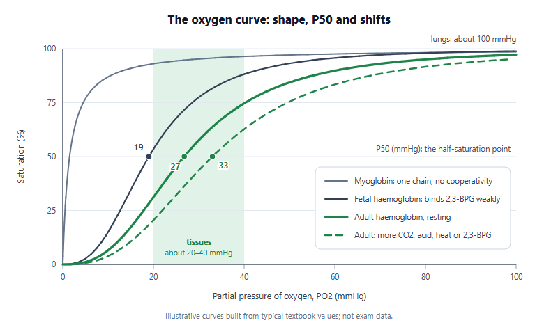
More carbon dioxide, more acidity (the Bohr effect), a higher temperature and more 2,3-bisphosphoglycerate (2,3-BPG) all shift the adult curve to the right. All four are marks of working tissue, so extra oxygen is released exactly where it is used. Further left means higher affinity. Fetal haemoglobin binds 2,3-BPG less strongly than adult haemoglobin, which lets it take oxygen from maternal blood, and myoglobin, a single chain without cooperativity, holds oxygen in muscle until PO2 falls very low. Comparative items reuse the logic: the llama and the bar-headed goose carry higher-affinity haemoglobin that loads well in thin air, while small mammals, with high metabolic rates, tend to have lower-affinity blood that unloads readily.
The hardest items separate saturation, the share of binding sites occupied, from content, the oxygen a volume of blood actually carries, which also depends on how much haemoglobin there is. Keep PO2, saturation and content in separate columns and most clinical stems resolve.
| Situation | Arterial PO2 | Saturation | O2 content | What the item is testing |
|---|---|---|---|---|
| Anaemia | Normal | Normal | Low | Too little haemoglobin: capacity falls while saturation stays normal |
| Carbon monoxide poisoning | Normal | Low: CO occupies binding sites | Low | CO binds roughly 200 times more tightly than oxygen and shifts the remaining curve left |
| Arriving at high altitude | Low | Low | Low | The gradient has fallen, not the oxygen percentage of the air |
| Blood leaving exercising muscle | Normal on the arterial side | Low in venous blood | Low in venous blood | Greater extraction, helped by a right shift from carbon dioxide, acid and heat |
| Cyanide poisoning | Normal | Normal; venous saturation stays unusually high | Normal | Delivery works, but mitochondria cannot use the oxygen |
Carbon dioxide travels mostly as bicarbonate, around 70% in typical textbook figures, formed by carbonic anhydrase inside red blood cells as chloride ions move in; roughly a fifth or more rides on haemoglobin, and a little stays dissolved. The Haldane effect partners the Bohr effect: deoxygenated haemoglobin carries more carbon dioxide, so unloading oxygen in the tissues helps blood collect carbon dioxide, and loading oxygen in the lungs helps it let go.
Pressure and flow: the cardiac cycle and the capillary bed
Heart valves are passive: they move only when the pressure on one side exceeds the pressure on the other. Track the pressures on the left side of the heart and the cycle writes itself.
- Ventricular pressure passes atrial pressure and the mitral valve closes: the first heart sound.
- With both valves shut, pressure rises at constant volume until it passes aortic pressure.
- The aortic valve opens and blood is ejected.
- As the ventricle relaxes below aortic pressure, the aortic valve closes: the second heart sound.
- Once ventricular pressure falls below atrial pressure, the mitral valve opens and filling resumes, topped up by atrial contraction.
Faults then become predictable. A narrowed aortic valve forces a higher ventricular pressure to eject the same volume, and a leaking mitral valve sends blood back into the atrium during contraction. Stroke volume is end-diastolic volume minus end-systolic volume; cardiac output is heart rate multiplied by stroke volume, and typical resting figures of about 70 millilitres and 70 beats a minute give roughly five litres a minute. On an ECG, the P wave is atrial depolarisation, the QRS complex is ventricular depolarisation (hiding atrial repolarisation) and the T wave is ventricular repolarisation. Much of the pause between P and QRS is the delay at the atrioventricular node, which lets the atria finish emptying.
In the vessels, flow equals the pressure difference divided by resistance, and for smooth flow resistance varies with the inverse fourth power of the radius: halve an arteriole’s radius and its resistance rises about sixteen-fold, which is why arterioles steer blood between organs. Blood moves most slowly in the capillaries because their combined cross-sectional area is the largest, not because each capillary is narrow. There, blood pressure pushes fluid out while plasma proteins draw water back in; in the classic textbook picture more fluid leaves than returns, and the lymphatic system collects the excess. Oedema items disturb that balance through low plasma protein, raised venous pressure or blocked lymph drainage.
Comparative designs: countercurrent gills, bird lungs and hearts
In a fish gill, water and blood flow in opposite directions, so at every point along the surface the water is still richer in oxygen than the blood beside it. A gradient survives along the whole length and blood leaves far better loaded than a same-direction design would allow; textbook accounts put the share of dissolved oxygen that gills remove from water at more than 80%.
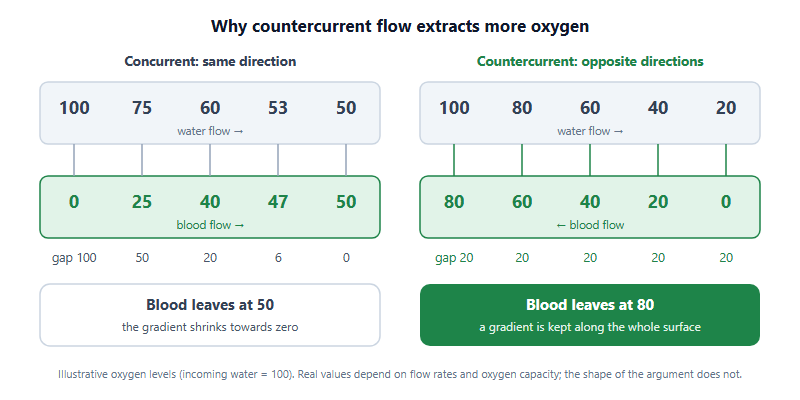
Watch the wording in stems. A countercurrent exchanger preserves a gradient passively, as in gills and in the heat-exchanging blood vessels in the limbs of many birds and mammals. The loop of Henle is a countercurrent multiplier, which spends energy on active transport to build a gradient. Options sometimes swap the two.
Birds use air sacs to keep air moving one way through the fine tubes of the lung, the parabronchi, during both inhalation and exhalation, so a single breath takes two breathing cycles to pass through. Insects pipe air straight to their tissues through tracheae, so their open circulatory system does little oxygen transport. Diving mammals such as seals store most of their oxygen in blood and myoglobin-rich muscle rather than in their lungs.
| Group | Heart | Circuits | What an item usually hinges on |
|---|---|---|---|
| Bony fish | Two main chambers: one atrium, one ventricle | Single: heart, gills, body | Pressure falls across the gill capillaries, so blood reaches the body at low pressure |
| Amphibians | Three chambers: two atria, one ventricle | Double: pulmocutaneous and systemic | Some mixing in the ventricle; the skin adds gas exchange |
| Non-crocodilian reptiles | Three chambers, ventricle partly divided | Double, able to shunt | Blood can bypass the lungs during diving or breath-holding |
| Crocodilians | Four chambers, with a connection between the two aortas | Double, with a possible shunt | Four chambers do not always mean zero mixing |
| Birds and mammals | Four chambers, evolved independently | Double, fully separated | High systemic pressure supports high metabolic rates; a classic convergence |
A two-week block, and the perturbation drill
This topic rewards a short, deliberate block more than long reading. One workable shape:
- Days 1–3: draw the adult curve from memory with P50 and both zones marked, then add fetal haemoglobin, myoglobin and a right shift, and explain each position in a sentence.
- Days 4–6: rebuild the saturation-versus-content table from a blank page and add two situations of your own.
- Days 7–10: sketch left ventricular and aortic pressure across one cycle, mark every valve event, and line the ECG waves up beneath it.
- Days 11–14: countercurrent exchange, bird lungs and heart designs, then timed mixed items.
Each day, run the perturbation drill: pick one variable, such as carbon dioxide, altitude, a valve, arteriole radius or plasma protein, and state what changes, in which direction, and what does not. That last clause matters, because many distractors describe a change that never happens. Then practise under time with our free past-paper pack, which gathers several years of papers, with worked solutions for some years rather than all. On the China route, the 2026 sitting was on 4 April; check the China-region page for the 2027 dates before you fix this block in your calendar.
Is circulation a separate section of the USABO?
No. It sits inside animal anatomy and physiology, which our own reading of the content makes the largest area. Check the current content outline published on cee.org.
What does a right shift of the oxygen curve mean?
Lower oxygen affinity at the same partial pressure, so haemoglobin unloads more readily. Carbon dioxide, acidity, heat and 2,3-BPG cause it.
Why does fetal haemoglobin sit to the left?
It binds 2,3-BPG less strongly than adult haemoglobin, so its oxygen affinity is higher and it can take oxygen from maternal blood.
Do I need to memorise blood pressure numbers?
Rarely as pure recall. Know typical values to sanity-check answers, but reason from pressure gradients and resistance instead.
This is an independent guide operated by Hanlin Education for China-based international-school students. We are not affiliated with, endorsed by, or sponsored by the Center for Excellence in Education (CEE). Exam format, content weightings, eligibility and dates are set by the organisers — please confirm current details on cee.org. Physiological values here are typical textbook figures, not exam data, and remarks on how questions are framed are our coaches' editorial judgement. If you spot an error, tell us and we will correct it within 7 working days.